42 neurons and supporting cells worksheet
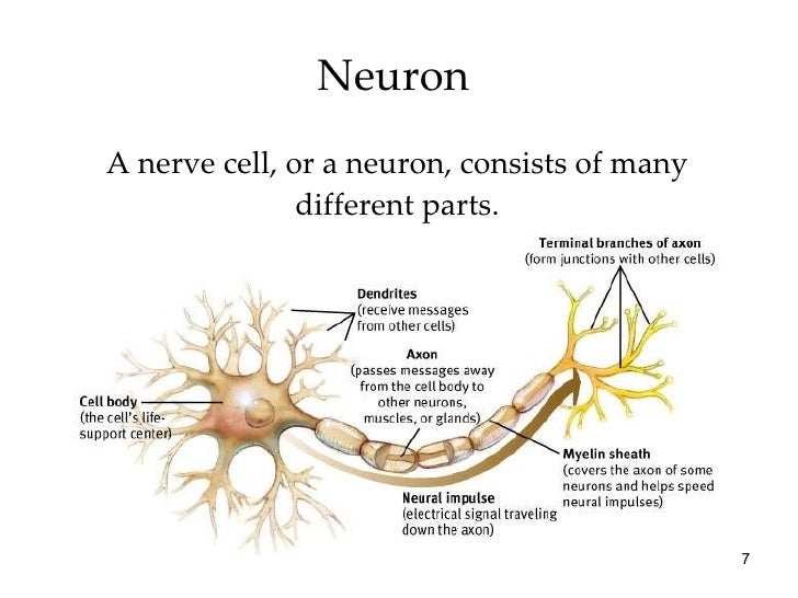
Anatomy of a neuron. Neurons, like other cells, have a cell body (called the soma ). The nucleus of the neuron is found in the soma. Neurons need to produce a lot of proteins, and most neuronal proteins are synthesized in the soma as well. Various processes (appendages or protrusions) extend from the cell body. Name four types of neuroglia in the CNS and list at least four functions of these cells. Neurons and supporting cells worksheet. 11 Neuroglia cells The function of neuroglia cells is to ensure structural support nourishment and neuron protection. 4What is the difference between neurons and neuroglia.
Students will learn about the 6 key features of the neuron and how neurons make connections with one another. This lesson includes interactive coloring and labeling neuron notes, a game with a baseball to simulate neurons making connections with one another, and an overall link to a growth mindset.
Neurons and supporting cells worksheet
Cells are densely packed and intertwined. There are two principal types of cells: neurons and supporting cells. In the following exercise, write . true. if the statement is true but if the statement is . false, change the underlined word to make the statement true and write the correct word on the answer blank. TRUE1. Supporting cells in the ... Billions of Cells: Type of Neurons" to answer the following questions: 1. ... The brain contains about 100 billion nerve cells (neurons) and trillions of support cells called __GLIA OR (GLIAL CELLS) ... (This worksheet was created by Joyce Taaffe, at teacher at Wheeler High School, Marietta, GA.) In the past, students would label a nerve cell and color neuroglia cells using paper handouts to learn the structures of the neuron and the neuroglia (supporting cells). I even have a neuron plushie to go along with this lesson and use toilet paper rolls to model the myelin sheath.
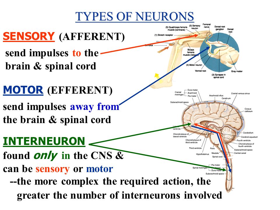
Neurons and supporting cells worksheet. cel IS are the supporting cells due to the length of its associated with axons, They form a(an) m around many vertebrate neurons, Nodes envier interrupt the myelin sheath were the axon is in direct contact with surrounding intercellular fluid, The junction between a neuron and a muscle is called ... cells, which can be classified into thousands of different. types. The two principal and distinct classes are nerve cells. (neurons) and neuroglial cells (or glia). The neuron is the. main ... The brain and nervous system are made of billions of nerve cells, called neurons. Neurons have three main parts: cell body, dendrites, and axon. The axon is covered by the myelin sheath. The transfer of information between neurons is called neurotransmission. This is how neurotransmission works: 1. Neurons: Neurons are the cells of the nervous system, which receive and transmit nerve impulses. Neuroglia: Neuroglia is the supporting nervous system cells, which provides mechanical and structural support to neurons. It supplies nutrients and oxygen to neurons, and supplies electrical insulation through axons of the neuron.
Transcribed image text: .. i MetroPCS令 9:28 PM ggc.view.usg.edu Worksheet 9 -NEURAL TISSUE&SPINAL CORD Important concepts of Neural Tissue: types of cells within the nervous system parts of a neuron the function of each part of the neuron different shapes of neurons (unipolar, bipolar, multipolar) types of supporting cells (neuroglia) and their function CELLS OF THE NERVOUS SYSTEM Cells of ... neurons. Supporting cells. are non-conducting cells that far outnumber the neurons and function to protect, support, and insulate the neurons. Neurons . are. the large conducting cells of nervous tissue. They all have a . nucleus-containing . cell . body, and their . cytoplasm. is drawn out into long extensions (glial cells) - Regulate the environment around the nucleus-Provide a supporting framework for neural tissue-Acts as phagocytes (engulf bacteria and pathogens) - Smaller than neurons but far outnumber them - Can reproduce, whereas neurons cannot. Supporting cells, called _____ are essential for the structural integrity of the nervous system and for the normal functioning of the neurons. Which glial cells provide structural and metabolic support for neurons? What do these glial cells also form? What cells form the insulating sheaths around axons? Where are these cells found? All cells ...
Supporting cells called neuroglia, small cells that surround and wrap the more delicate neurons Neurons, nerve cells that are excitable (responsive to stimuli) and transmit electrical signals Figure 4.10 on p. 129 will refresh your memory about these two kinds of cells before we explore them further in the next two modules. 1. Nerve tissue is composed of 2 main types of cells: Neurons-nerve cells that are specialized to detect and react to stimuli, by generating and conducting nerve impulses. Neuroglial cells-accessory cells for filling spaces and supporting neurons. 2. Microscopic anatomy of neurons All neurons have a cell body called soma which contains a Chapter 12 Nervous Tissue 12 1 Ch 12 from slidetodoc.com The nervous system is composed of excitable nerve cells (neurons) and Neurons the central nervous system is composed of what organs? The nervous system lesson includes a powerpoint, student guided notes, teacher notes, and a two page worksheet with answers. Synapses: Send electrical impulses to neighboring neurons. 2. Myelin sheaths: Cover the axon and work like insulation to help keep electrical signals inside the cell, which allows them to move more quickly. 3. Axon: Transfers electrical impulse signals from the cell body to the synapse. 4. Soma: The cell body which contains most of the cell’s ...
Neuroglia provide important support for nerve cells and this quiz/worksheet combo will test your understanding of their characteristics. Some things you'll be assessed on include the names and ...
The nervous system is made up of neurons, specialized cells that can receive and transmit chemical or electrical signals, and glia, cells that provide support functions for the neurons by playing an information processing role that is complementary to neurons.A neuron can be compared to an electrical wire—it transmits a signal from one place to another.
This 6-page worksheet product (with 2 pages of answer keys) is designed to introduce upper middle school and high school anatomy students to the nervous system, neurons, nerves, and glial cells. A 4-page reading section covers the following topics: functions of the nervous system, CNS vs PNS, neuron
The cell and science of a motor neuron or tissue? Name four types of neuroglia in the CNS, and even movement. He spends plenty of nervous and worksheet: neurons and a cyst in. Nerves that carry out basic graph with other by physical is a baseball pitched to communicate with epilepsy to accumulate and spinal cord to design drugs are still very similar.
Neuroglia are support cells that perform a variety of functions. Neuroglia cells account for over half the brain's weight. There can be 10 to 50 times more neuroglia cells than neurons in various locations in the brain. Neurons FIGURE 11.4 1. The neuron cell body contains the nucleus, and the cell body is the site of manufacture of
Cells of the epithelial tissue was different shapes as shown on the student's worksheet Cells can be. Seen in pictures of small brain as well while some other structures under it. Structural Classification Functional Classification Nervous Tissue Structure and Function Supporting Cells Neurons Central Nervous System.
Nervous System Worksheet Answers. 1. The diagram below is of a nerve cell or neurone. i. Add the following labels to the diagram. ii. If you like, colour in the diagram as suggested below. iii. Now indicate the direction that the nerve impulse travels.
Which glial cells provide structural and metabolic support for neurons, and also help to maintain the blood-brain barrier? What two cells form the insulating sheaths around axons? Where are each of these cell types found? All cells have an electrical charge difference across their plasma membrane called the _____.
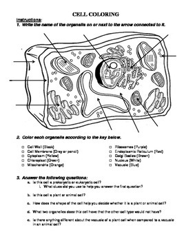
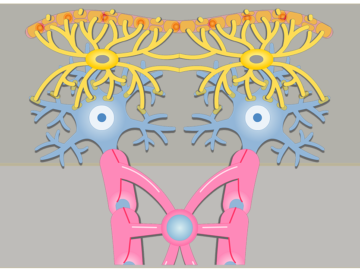
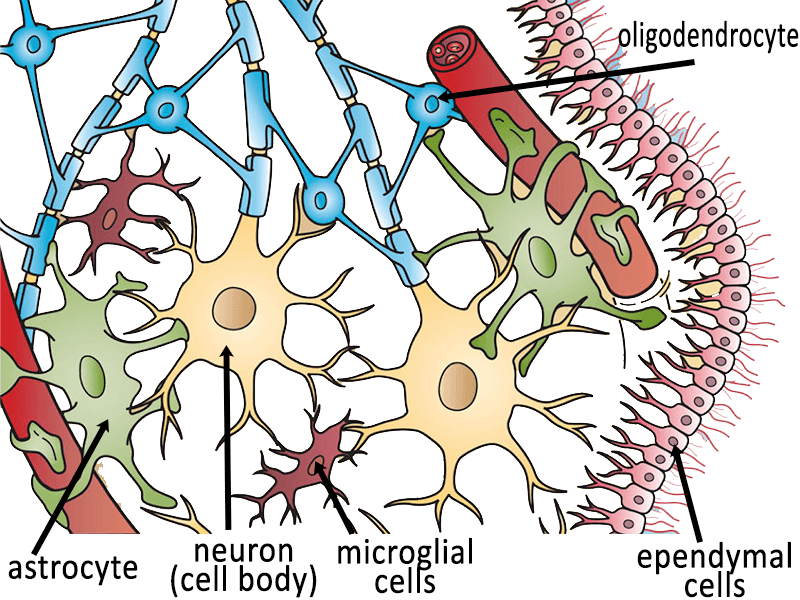
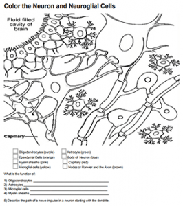
Nerve Cells Coloring. KEy. Oligodendrocytes (purple) Astrocyte (green) Body of Neuron (blue) Ependymal Cells (orange) For each of the cells above, color the nucleus a darker shade of purple, green, blue, orange. Myelin sheaths (pink) Capillary (red)
Start studying Neuroglia (Glial cells) = Supporting Cells. Learn vocabulary, terms, and more with flashcards, games, and other study tools. Search. Create. ... a protective barrier or mechanism that helps maintain a stable environment for the neurons of the brain. Ependymal Cells. Line cavities of brain and spinal cord. Ependymal Cells. Many ...
They transmit the information to other neurons and/or other kinds of cells (such as muscle). A typical neuron has an enlarged area, the cell body, which contains the nucleus. Neurons typically also have two types of specialized extensions that project away from the cell body. The branches on which information is received are known as dendrites.
In the past, students would label a nerve cell and color neuroglia cells using paper handouts to learn the structures of the neuron and the neuroglia (supporting cells). I even have a neuron plushie to go along with this lesson and use toilet paper rolls to model the myelin sheath.
Billions of Cells: Type of Neurons" to answer the following questions: 1. ... The brain contains about 100 billion nerve cells (neurons) and trillions of support cells called __GLIA OR (GLIAL CELLS) ... (This worksheet was created by Joyce Taaffe, at teacher at Wheeler High School, Marietta, GA.)
Cells are densely packed and intertwined. There are two principal types of cells: neurons and supporting cells. In the following exercise, write . true. if the statement is true but if the statement is . false, change the underlined word to make the statement true and write the correct word on the answer blank. TRUE1. Supporting cells in the ...
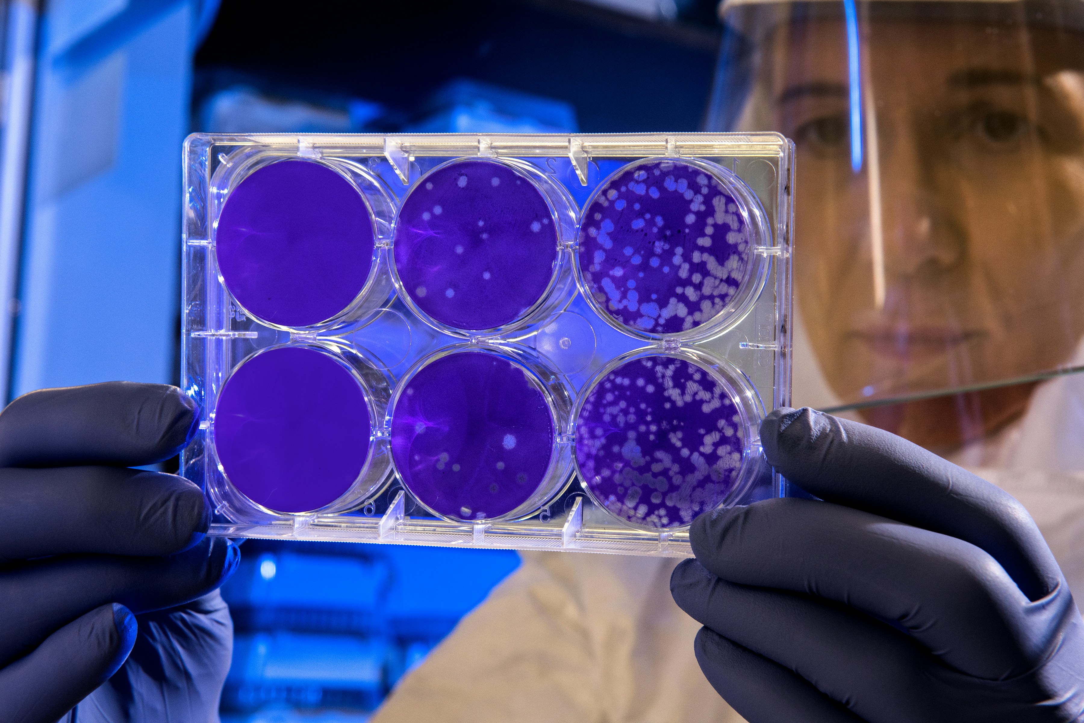
Scientist examines the result of a plaque assay, which is a test that allows scientists to count how many flu virus particles (virions) are in a mixture. To perform the test, scientists must first grow host cells that attach to the bottom of the plate, and then add viruses to each well so that the attached cells may be infected. After staining the uninfected cells purple, the scientist can count the clear spots on the plate, each representing a single virus particle.

Drosophila Wing Cells. The human CBFA2T3-GLIS2 fusion protein is a key driver of pediatric acute megakaryoblastic leukemia (AMKL), and confers a poor prognosis. Researchers found a way to express CBFA2T3-GLIS2 (red) in larval Drosophila (fruit fly) wing disc cells, confirming a major role for the BMP signaling pathway. This pathway may provide a target for new therapies. Nuclei (green) and actin filaments (purple) are also shown.
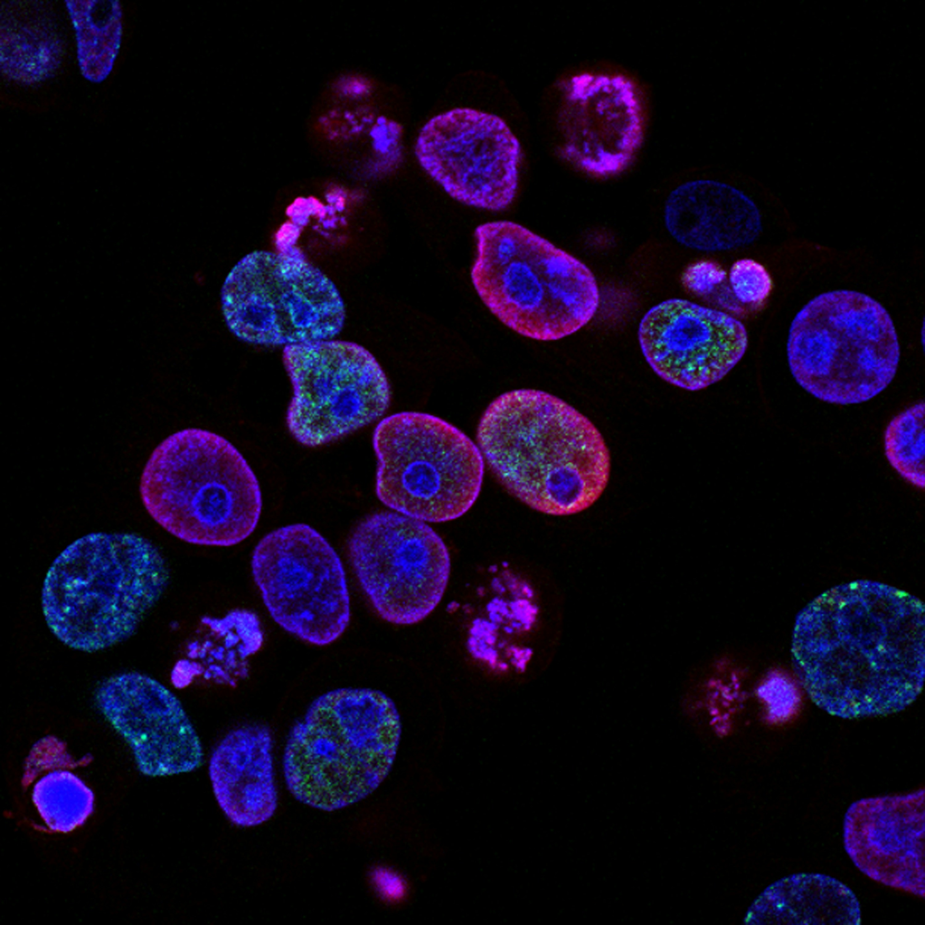
Human colorectal cancer cells treated with a topoisomerase inhibitor and an inhibitor of the protein kinase ATR (ataxia telangiectasia and Rad3 related), a drug combination under study as a cancer therapy. Cell nuclei are stained blue; the chromosomal protein histone gamma-H2AX marks DNA damage in red and foci of DNA replication in green. Created by Yves Pommier, Rozenn Josse, 2014




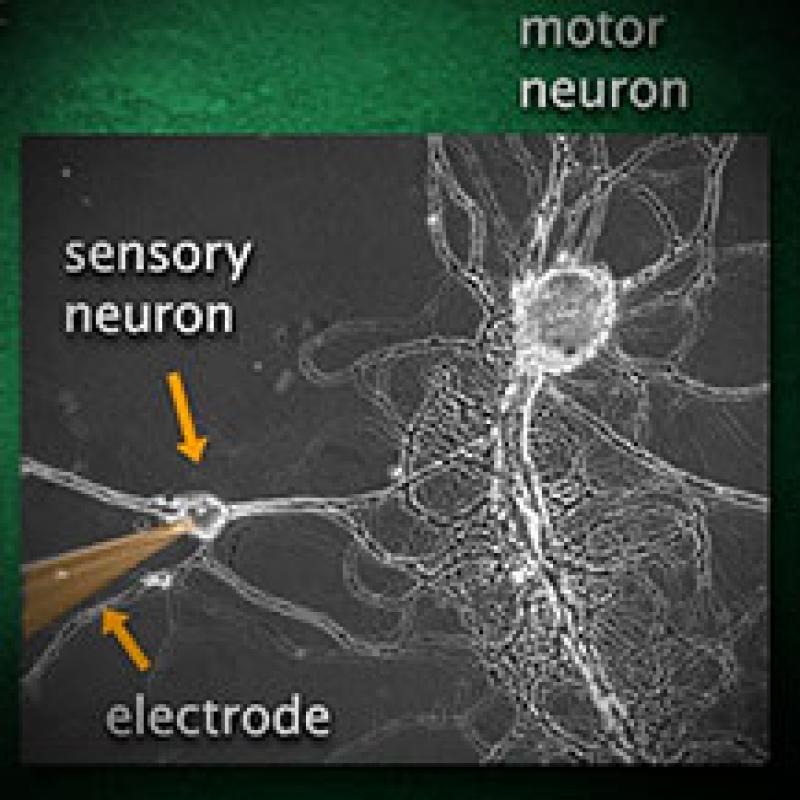






:background_color(FFFFFF):format(jpeg)/images/library/10996/anatomy-neurons-basic-types_english.jpg)




0 Response to "42 neurons and supporting cells worksheet"
Post a Comment